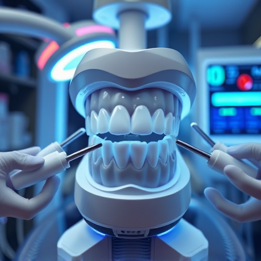


Intraoral cameras transform dental examinations by providing high-resolution images of the oral cavity, aiding in early detection of tooth decay and gum disease. A meticulous preparation process ensures a comfortable examination. The camera captures detailed mouth images from all angles, transmitting them to a monitor for both patient and dentist viewing. These visual aids enhance diagnostic accuracy, enabling early identification of issues like decay or gum disease through precise zooming.
Discover the power of intraoral camera examinations—a revolutionary tool in dental care. This non-invasive, high-tech approach enhances traditional visual inspections, allowing dentists to detect issues early. From understanding the technology to preparing for the exam and capturing detailed images, this step-by-step guide demystifies the process. Learn how intraoral cameras provide a clear view of oral health, enabling accurate diagnoses and effective treatment planning, all while making dental visits more comfortable for patients.
- Understanding Intraoral Cameras: Tools for Visual Inspection
- Step-by-Step Preparation for an Effective Exam
- Capturing and Analyzing Images During the Examination
Understanding Intraoral Cameras: Tools for Visual Inspection

Intraoral cameras are revolutionary tools that have transformed the way dental professionals conduct examinations and provide comprehensive dental care. These advanced devices enable dentists to capture detailed, high-resolution images of a patient’s oral cavity, offering a clear and precise view of teeth, gums, and other structures within the mouth. This technology is particularly beneficial for detecting early signs of tooth decay, gum disease, or abnormalities that might be invisible during traditional visual inspections.
By integrating intraoral camera examination into their practices, dentists can offer more efficient and accurate diagnoses. The cameras facilitate discussions with patients about their oral health, as the visuals provide a shared understanding of any issues identified. Moreover, these tools are not limited to diagnosis; they also play a role in treatment planning, especially for procedures like dental bonding or teeth cleaning, ensuring that every aspect of oral care is thoroughly assessed and addressed.
Step-by-Step Preparation for an Effective Exam
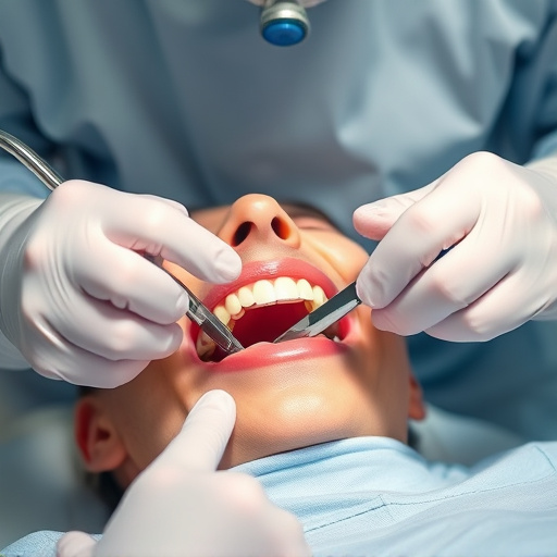
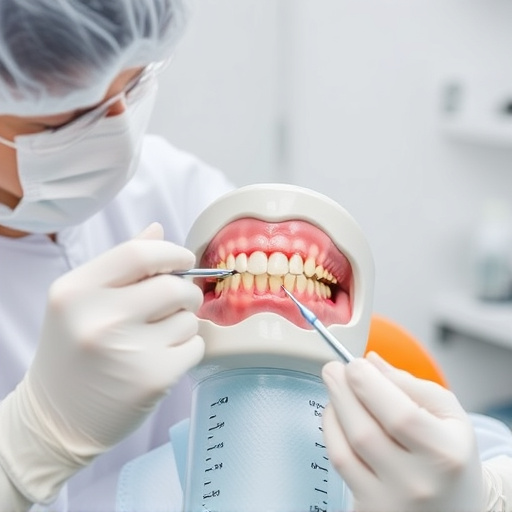
Before conducting an intraoral camera examination, a thorough preparation process is essential to ensure an effective and comfortable experience for both the patient and dentist. The first step involves gathering all necessary equipment, including the intraoral camera, lighting sources, and a computer monitor. It’s crucial to verify that the camera is properly calibrated and the software is updated to guarantee accurate and high-quality images.
Additionally, proper patient preparation is key. Communicate with your patient about the procedure, explaining what to expect during the exam. Encourage them to express any concerns or questions they may have. From a dental perspective, consider the patient’s oral health history and current conditions, such as tooth decay, gum disease, or existing dental crowns. This step allows for a more comprehensive examination, focusing on both preventive dentistry and identifying potential issues that may require immediate attention.
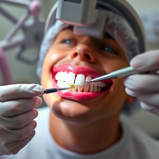
Capturing and Analyzing Images During the Examination
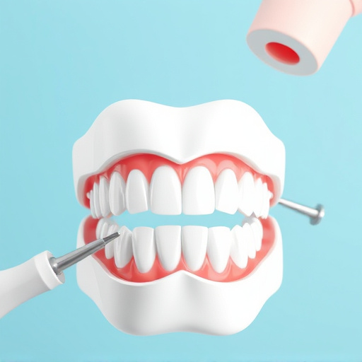
During an intraoral camera examination, the dental professional uses a tiny, high-definition camera to capture detailed images of your mouth – from every angle. This innovative technology allows for a more comprehensive visual assessment compared to traditional methods. The camera is typically attached to a small handle or wand and can be easily maneuvered inside your mouth. As the camera captures images, it transmits them in real-time onto a monitor outside, allowing both you and your dentist to see up-close what’s happening within your oral cavity.
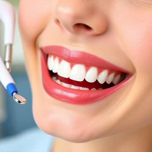
These captured images are not just for visual reference; they play a crucial role in diagnostic accuracy. Using specialized software, the dentist can analyze these intraoral camera images to identify subtle issues like tooth decay, gum disease, or even potential problems with dental implants or children’s dentistry needs. By zooming in on specific areas and capturing close-ups, the technology enables precise identification of caries, cracks, or inflammation – often much earlier than traditional methods can detect.
An intraoral camera examination offers a comprehensive, non-invasive view of your oral health. By following these simple steps, from understanding the technology to capturing detailed images, you can ensure a thorough assessment. This advanced tool allows for early detection of issues, enabling prompt treatment and maintaining optimal oral well-being. Embrace this modern approach to dentistry and take control of your dental care with the power of intraoral camera examinations.